Total Hip Replacement Surgery
Total Hip Replacement Surgery was first performed in 1960, almost 60 years ago. Since then, materials, technology and surgical techniques have greatly increased the effectiveness of total hip replacement. Over 300,000 hip replacements are performed each year in the U.S. Hip replacement surgery is one of the most successful operations in all of medicine.
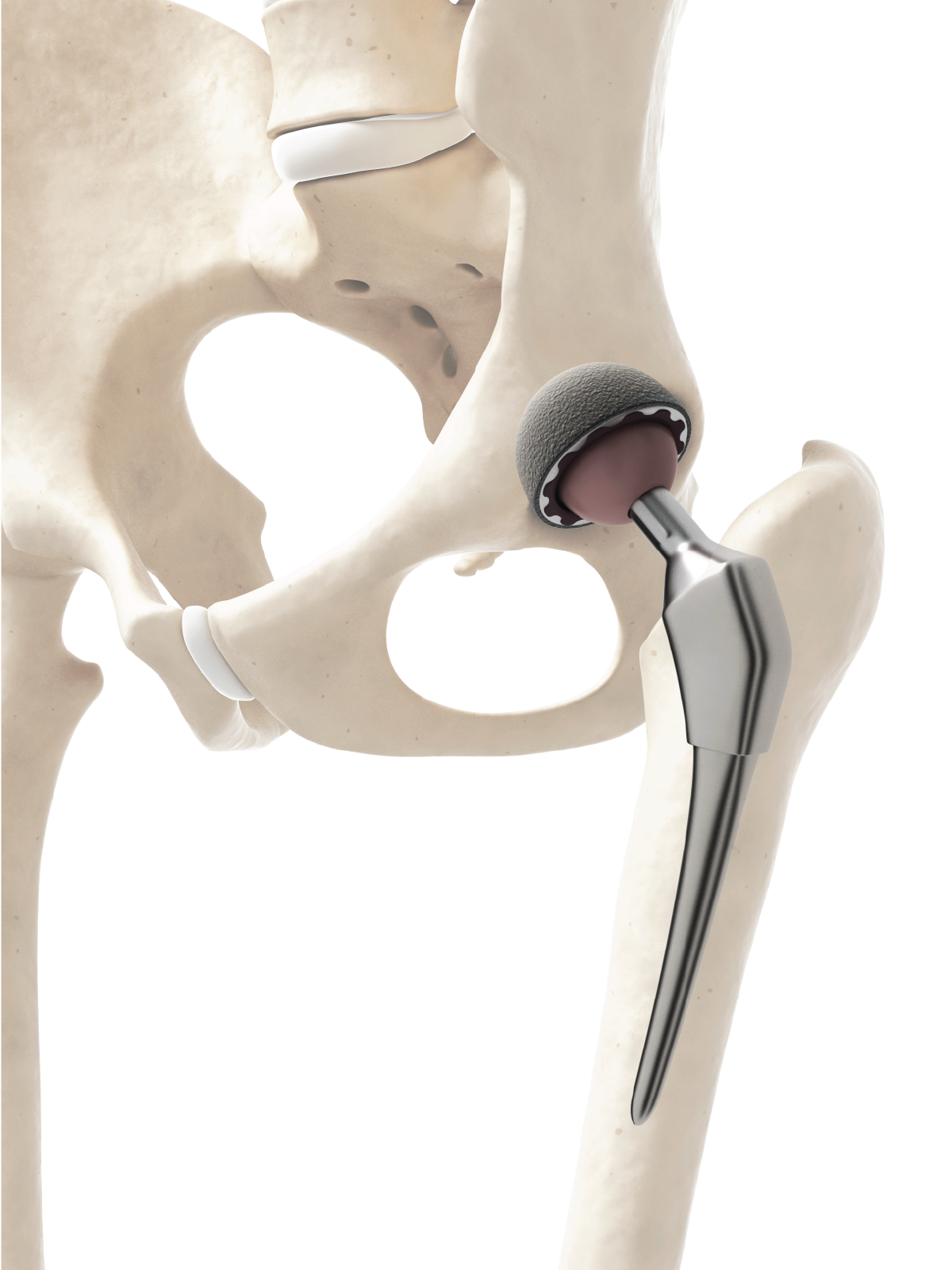
The hip is one of the body’s largest joints and allows for a tremendous range of movement. Total hip replacement surgery consists of a specially designed ball and socket to replace your damaged or diseased hip joint. The ball and stem replace the worn ball at the end of your femur. A cup replaces the rough hip socket in your pelvis. The implant has smooth surfaces that fit together to insure that the ball moves easily and painlessly within the socket as it would in a healthy hip.
Anterior Supine Intramuscular Approach
Dr. Walter performs a muscle-sparing, direct anterior, minimally invasive, total hip technique rather than the much more intensive traditional hip replacement still performed by most surgeons. Dr. Walter brought this procedure to the region 13 years ago and has successfully performed more than 2000 surgeries using this technique. This approach is anterior and through a much smaller incision. This preserves the muscle and reduces trauma to the surrounding tissues resulting in less pain and a faster return to normal activity. Most of Dr. Walter’s patients walk within hours after surgery.
Out-patient Total Joint Surgery
Joint replacement surgery has undergone tremendous evolution over the past 20 years. With improved techniques, post-operative pain management and prosthetic designs we are now able to offer out-patient total joint replacement surgeries on select candidates. If you are interested in exploring this option further, please discuss this with Dr. Walter at your next visit.
Hanna Orthopedic Table
Dr. Walter performs most hip replacement surgeries using the Hana Orthopedic Table. The Hana table is specifically designed for the Direct Anterior Total Hip Arthroplasty. The Hana table allows the surgeon to approach the hip comfortably through the front with a small incision. This muscle sparing technique is less invasive than traditional methods. Live x-ray imaging is used during surgery to allow for optimal component position.
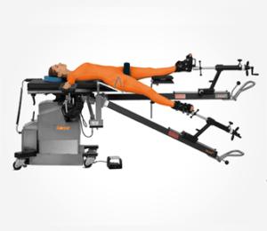
Hanna Orthopedic Table
Related Videos
Shoulder Replacement Surgery
There are two types of shoulder replacement surgery: Total Shoulder Arthroplasty and Reverse Total Shoulder Arthroplasty. The standard Total Shoulder Arthroplasty is designed for patients with an intact rotator cuff. A Reverse Total Shoulder Arthroplasty is for patients with chronic rotator cuff dysfunction.
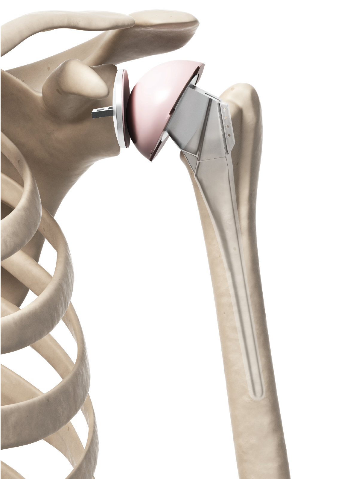
The shoulder joint is a ball and socket joint. The ball is the humerus, (the top of the arm bone) while the socket lies within the shoulder blade (scapula). This joint is vital to everyday activity as it allows for a tremendous range of movement. During shoulder replacement surgery, the ball is removed from the humerus and replaced with a shaped metal implant. The implant is attached to the humerus. The socket is shaved clean and replaced with a new plastic socket fitted into the scapula to enable the joint to move smoothly without pain. Physical Therapy is critical to the success of a shoulder replacement and should be taken seriously.
Knee Replacement Surgery
Knee replacement surgery was first performed in 1968. Since then, techniques and materials have improved dramatically making total knee replacements one of the most successful procedures in medicine today with more than 600,000 knee replacements performed each year in the U.S. and projected to increase 186% by 2030.
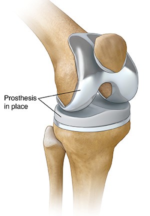
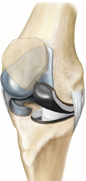
Total Knee Replacement
Total Knee Replacement surgery addresses damage to the knee as a result of arthritis or injury. This involves removing parts of the damaged surface and replacing them with a smooth metal surface. There are three parts of any knee replacement:
- Femoral component: This is a contoured metal piece that fits over the end of the femur and allows the knee to bend and straighten smoothly.
- Tibial component: A flat metal and polyethylene plate that covers the top of the tibia.
- Patellar component: This dome shaped plastic component allows the kneecap to glide smoothly over the new knee.
Partial Knee Replacement
A Partial Knee Replacement or “Uni” may be performed when only a portion (one compartment) of the knee is damaged and needs replaced. There are three compartments of the knee but 90% of partial knee replacement patients only require the medial compartment to be replaced. A partial knee replacement is performed through a smaller incision and can often done as an out-patient procedure.